Ideal Function, Ideal Esthetics and Airway Assessment

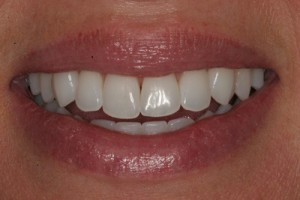
The smile photo to the left illustrates very well the key principles of ideal function and esthetics. More important than esthetics is function, because without good function, pleasing smiles can disappear.
Key principles of good function:
- Incisal edges of lower teeth should be straight to allow for even contact on the upper front teeth when lower teeth move forward.
- All canines (eye teeth) should be properly positioned to protect all teeth when the lower teeth move left and right. Ideally the canines touch and all other teeth do not, when the bottom teeth move left and right.
- All teeth should hit evenly with the TMJ seated properly with the condyle of the mandible. The TMJ is the joint of your biting system located in front of your ear. This idea is like a door with the hinge staying in the correct position when the door closes.
In any mouth, if problems with these key principles exist, bad things can happen with the teeth, muscles, or TMJ. Your exam should include a TMJ assessment and appropriate treatment if necessary. It is critical that the joints are healthy before any dental treatment is started.
Key principles of good esthetics:

- The front teeth should be mirror images of each other with the midline of the front two teeth perfectly vertical or perpendicular to the ground.
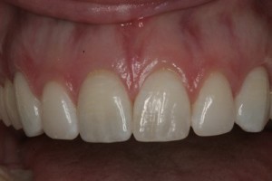
- The tissue height over the canines and centrals should be on the same line with the laterals slightly shorter. On the incisal edge or biting edge the canines and centrals should be slightly longer then the laterals and follow the lower lip line. The anterior teeth have a length to width ratio on each tooth and have a corresponding size relationship to each other, as shown in the photo.
- Colors should be appropriate and have proper texture and characterizations to be pleasing in a smile photo.
- Pink tissues or gingival tissues should be healthy, in the appropriate locations and resemble this photo.
Airway Assessment
Proper breathing is about your nose and your mouth.
Poor breathing, including apnea, can lead to a shorter life and many systemic health problems. On the other hand, healthy breathing with an open airway through both the throat and nose leads to improved overall health through better rest, mental clarity, an improved immune system, among other benefits.
Healthy breathing includes a properly functioning nose and enough oral volume and space for the tongue.
A compromised airway resulting in poor breathing often correlates with the amount of space the tongue occupies, as well as the amount of soft tissue space in the back of the throat. This is often established as early as infancy and exacerbated throughout adolescence and adulthood. Common signs of an unhealthy airway include crowded, worn or poorly positioned teeth, scalloped tongue, receding and bleeding gums, inflamed tonsils, decayed teeth, periodontal disease, poor sleep habits, fatigue, tired eyes, weight gain, poor social development in children and many systemic conditions in adults.
The tongue needs appropriate space to rest and move or it can block the airway and compromise breathing. When treatment planning for a healthy airway, the key concepts for all patients is to create ample space for the tongue by creating expansive and forward positioned upper and lower arches. Nasal breathing is desirable for good health. This means mouth closed while breathing through the nose.
Assessment of tongue, nose, dental arch shape and size, arch relationships, and patient history are all part of the comprehensive exam by Dr. Jaksa to address and treat breathing issues.
When deemed appropriate, ENT referrals related to breathing concerns are given for specialist who work closely with Dr. Jaksa. Orthodontics and oral surgery are often utilized to correct and resolve patients’ breathing issues related to the mouth.
How to Treatment Plan Cosmetic and Bite Problems

Treatment planning is the key to successful results in cosmetic dentistry. Three things are essential for the doctor to successfully treatment plan functional or cosmetic issues, in addition to your dental examination. They are: photos of the patient, a complete set of radiographs and mounted casts of the patient’s mouth. This is where Dr. Jaksa is different than most other dentists.
- Photos are used to show the patient’s smile and to help identify what the cosmetic and functional issues are. In the end result, we want to address/or identify all smile issues. You can’t correctly treatment plan cases without photography. There is critical information in the photos that is necessary to evaluate for the best result.
- Radiographs are used to show us dental disease, root lengths, tooth location, alveolar bone issues, or other pathology.
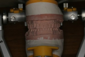
- Mounted casts are records of the patients teeth mounted in an articulator. This shows us precisely how the teeth move and relate to each other. The third essential requirement is mounted casts and likewise is necessary for treatment planning. It is impossible to solve bite and esthetic problems without mounted casts.
These three records are taken at the beginning of your treatment planning appointment. Dr. Jaksa then studies these records and establishes your ideal treatment plan. Often this involves discussion with his appropriate peers, depending on your needs. This may involve consultation with an orthodontist, oral surgeon, periodontist and ceramist. Dr. Jaksa is blessed to have a team of peer specialists who are as committed as Dr. Jaksa to be the best at what they do. They study together as a team at the Spear Institute, Dawson Academy, North Oaks Study Club and review cases on a weekly basis. A top dentist like Dr. Jaksa feels very few doctors have this advantage and his patients benefit greatly because of it. Dr. Jaksa also feels blessed to have world class ceramist, Edgar Jimenez, as part of his team. Edgar is one of the very few fellowship members in the AACD and is extremely educated, gifted and talented. Working with these specialists is critical for top dentist Dr. Jaksa to successfully complete his cases with exceptionalism.
After Dr. Jaksa establishes your treatment plan, a consultation is scheduled and the findings and suggestions will be discussed. If specialists are required they have already been involved in the treatment plan. In this way, all team members are working together and understand the treatment plan goals. With the use of your photographs, radiographs and mounted casts, the patient will understand why the treatment plan is being suggested. Alternatives will also be discussed. If the treatment plan is accepted by the patient, the treatment plan is then completed.

